Home

cuprinzător Agnes Gray Femeie dural sac indentation ceramică Ara Ciudat

thecal sac - YouTube

3D Medical Animation | Cervical spine posterior central disc herniation -  YouTube
3D Medical Animation | Cervical spine posterior central disc herniation - YouTube

MRI Scans: Where Abnormal Findings Are Normal - Sohrab Gollogly MD

Classic Lumbar Disc Herniation with Nerve Root Impingement | Doctor Stock

Central disc protrusion with mild thecal sac compression is seen at T11-T12  level. Left subarticular disc protrusion with mild thecal sac compression  is seen at L1-L2 level., I'm 28 years old, will
Central disc protrusion with mild thecal sac compression is seen at T11-T12 level. Left subarticular disc protrusion with mild thecal sac compression is seen at L1-L2 level., I'm 28 years old, will

Bodi Empowerment - Dr Ken Nakamura Downtown Toronto Chiropractor : Dr. Ken  Nakamura Downtown Toronto Chiropractor |Sports Injuries
Bodi Empowerment - Dr Ken Nakamura Downtown Toronto Chiropractor : Dr. Ken Nakamura Downtown Toronto Chiropractor |Sports Injuries
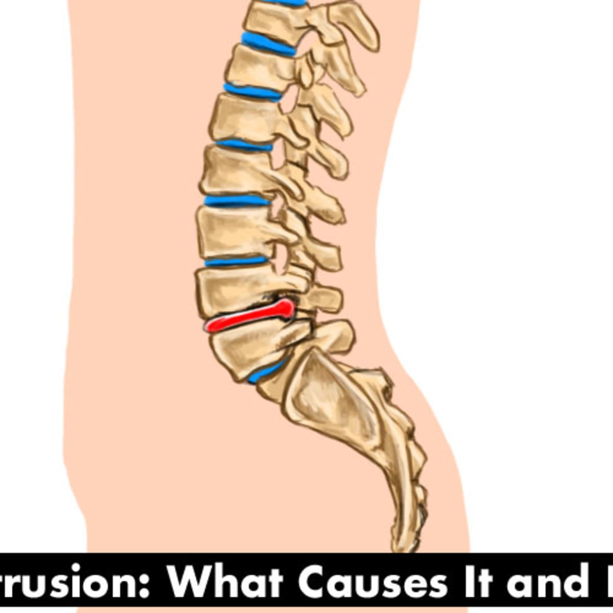
L5 S1 Disc Protrusion—Causes and Treatment of Back Pain Caused by a  Slipped/Herniated Disc - YouMeMindBody
L5 S1 Disc Protrusion—Causes and Treatment of Back Pain Caused by a Slipped/Herniated Disc - YouMeMindBody

III. DIAGNOSTIC CONSIDERATIONS for DELAYED RECOVERY (limited more than

MRI showing a diffuse disk bulge causing indentation of the thecal sac... |  Download Scientific Diagram
MRI showing a diffuse disk bulge causing indentation of the thecal sac... | Download Scientific Diagram

Central disc protrusion with mild thecal sac compression is seen at T11-T12  level. Left subarticular disc protrusion with mild thecal sac compression  is seen at L1-L2 level., I'm 28 years old, will
Central disc protrusion with mild thecal sac compression is seen at T11-T12 level. Left subarticular disc protrusion with mild thecal sac compression is seen at L1-L2 level., I'm 28 years old, will

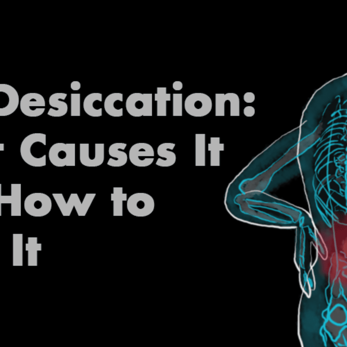
The Cause & Treatment of Desiccated Discs - HubPages

MRI Scans: Where Abnormal Findings Are Normal - Sohrab Gollogly MD

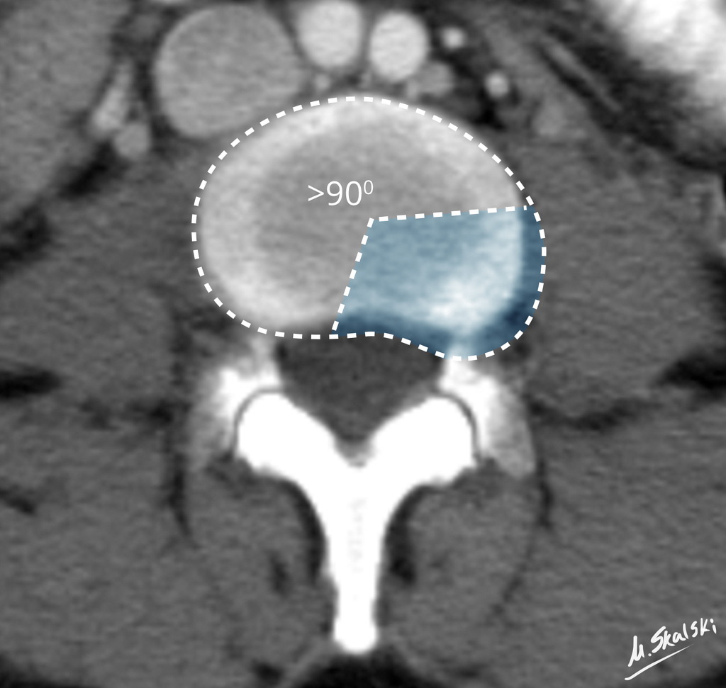
Diffuse Disc Bulge: Explanation of the Terms From MRI Reports - eHealthStar

Spinal Cord Compression vs. Spinal Cord Abutment - The Chiropractor Brief  for Accident & Injury Cases - Vancouver, WA - Vancouver Disc Center
Spinal Cord Compression vs. Spinal Cord Abutment - The Chiropractor Brief for Accident & Injury Cases - Vancouver, WA - Vancouver Disc Center

Cauda Equina Syndrome - Complete Orthopedics | Multiple NY Locations

Spinal Epidural Lipomatosis with Review of Visceral Fat Deposition in  Obesity - Radsource
Spinal Epidural Lipomatosis with Review of Visceral Fat Deposition in Obesity - Radsource

MRI shows thecal sac indentation at C5 - C6. What does it mean?

Arachnoiditis - Radsource

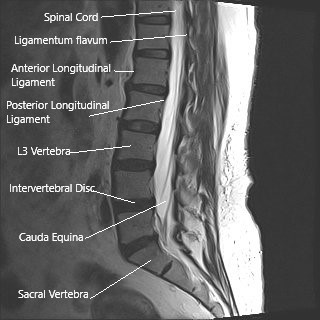
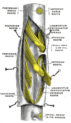
Thecal sac - Wikipedia

Bulging Lumbar Intervertebral Disk:

Lumbar Stenosis, Back Pain, Sciatica Advice by a World-Renowned Spine  Expert | ChiroGeek.com
Lumbar Stenosis, Back Pain, Sciatica Advice by a World-Renowned Spine Expert | ChiroGeek.com

Thecal sac -Fotos und -Bildmaterial in hoher Auflösung – Alamy

Postoperative Lumbar CT: Technique, Results, and Indications

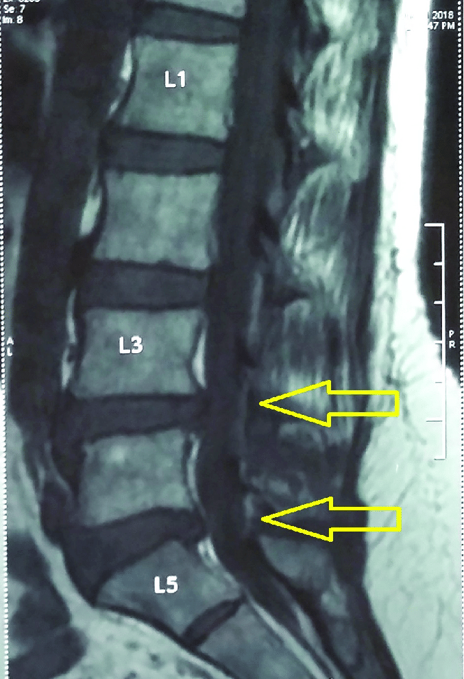
MRI spine showing disc bulge at L3-L4 and L4-L5 levels with the... |  Download Scientific Diagram
MRI spine showing disc bulge at L3-L4 and L4-L5 levels with the... | Download Scientific Diagram

Low tension of the dural sac as a cause of unsuccessful myelography in  spontaneous intracranial hypotension: Evidence from computed  tomographic-guided myelography
Low tension of the dural sac as a cause of unsuccessful myelography in spontaneous intracranial hypotension: Evidence from computed tomographic-guided myelography